Playback speed
10 seconds
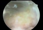
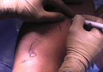
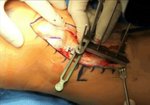
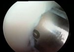
Tibial Tubercle Osteotomy and DeNovo Cartilage Transplant for Patellar Chondral Defect
779 views
June 23, 2016
This revised video is in response to the following comments:
Alan Merchant 1 week, 2 ...
read more ↘ days ago
I and several colleagues have watched your video presentation. From the information you have provided, none of us can understand how moving the tibial tubercle medially and releasing the lateral retinaculum can reduce pressure on a medial patellar lesion.
Is there more information that was not presented that justifies this approach?
Could you please help us understand why you chose these two surgical techniques, especially in a 14 year old girl?
Sincerely, Al Merchant
Laith Jazrawi 1 week, 1 day ago
Thanks for the comment Alan
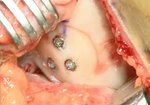
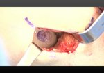
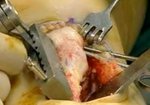
The title is misleading as the lesion you see from the video is more centrodistally based. 60 degree cut was utllized to provide more of an anteriorization and less medialization. The lateral release was performed as she did have exquisite lateral facet pain, +lateral compression pain and lateral tilt. The mri cut is misleading in terms of lesion location as the actual lesion from the open surgery shows its true location. We will modify title and put better MRI images. Thanks for comments. Sorry for confusion.
Laith M.Jazrawi MD
Alan Merchant 6 days, 15 hours ago
Dear Dr. Zazwari,
I assumed Dr. Capo was the surgeon. Were you the surgeon or the assistant or the consultant?
You have not changed the Video Title, just the announcement of the video on VuMedi's site. And there are no "better MRI images" as promised.
The history presented only medial pain and no mention of the "exquisite lateral facet pain".
The information presented in the video leaves one with the distinct impression that the patient's medial knee pain and medial MRI cartilage lesion was justification for both the AMZ and lateral release. And the true central location of the "medial lesion" was only recognized at the time of the arthrotomy.
Because of this unresolved confusion, the video itself loses all value as a teaching aid, and in fact teaches the wrong message.
Thank you for your attention,
Dr. Alan Merchant
Laith Jazrawi 8 minutes ago
Dr. Merchant we have clarified the video based on your recommendations. The plan was indeed initially a straight anteriorization but the near central location of the lesion found at the time of arthrotomy made us more comfortable with the 60 degree AMZ. She also had mostly lateral pain which was not included in our intro slides. This made us more comfortable performing the AMZ. The presentation is better with your input.
↖ read less
Login to view comments.
Click here to Login


